Last week I posted about my 4 year old BO hen with crop stasis. I treated her twice daily by removing material from her crop with a red rubber feeding tube and flushing crop, giving her antifungal and antibiotic therapy, and feeding her yogurt via the crop tube. She also got subcutaneous fluids twice a day and wore a crop bra. Both Sunday and Monday, I was unable to aspirate any material from the crop, so monday afternoon I anesthetized her and did a mini-exploratory on the crop. I removed a LOT of grain and grass from the crop, flushed it and recovered her, but was not hopeful as I felt no obvious obstruction. This morning her crop was full of fluid again. I euthanized and necropsied her this afternoon.
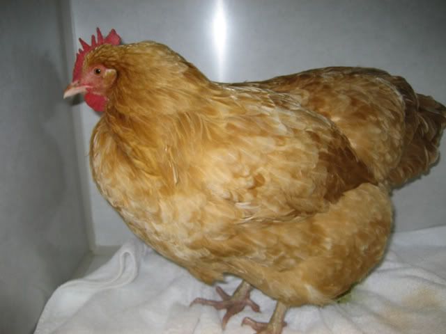
Here is a little tour of the chicken digestive tract and her disease, starting with a picture of Ivy in the clinic.
Ivy in her kennel.
Now for the necropsy pictures (graphic)... I made them big so you could see better detail. I started at her beak and cut down through the digestive tract until I found the problem.
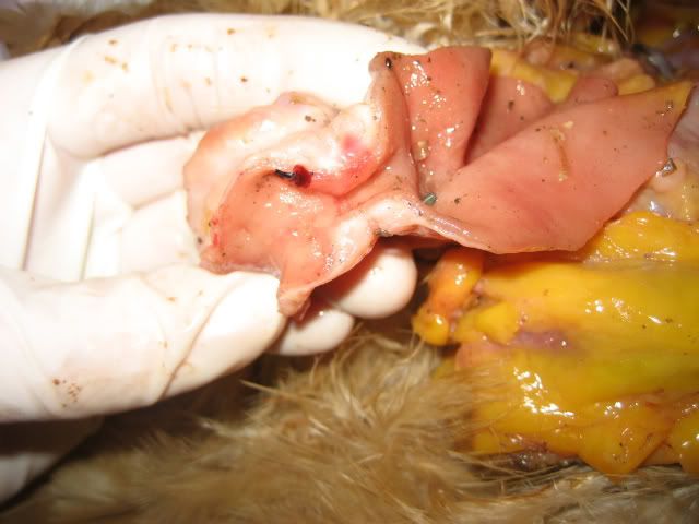
Crop, the surface looks healthy and there is no evidence of fungal infection. It was full of greenish/yellow fluid. The red is a postmortem change, it was not like that when I first opened her up.
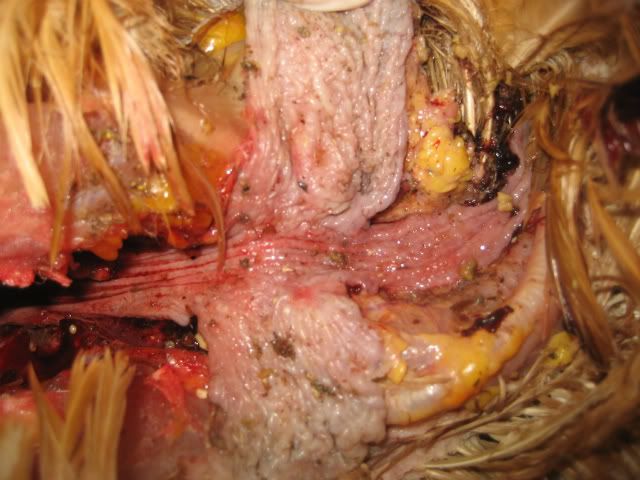
The glandular stomach (proventriculus) is on the right, the gizzard is on the left. Both were packed full of seeds, grains, little rocks, grass, layer pellet material. Again, the red in the proventriculus is a postmortem change.
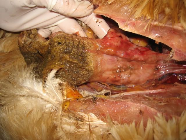
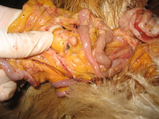
This is the intestine just after it left the gizzard. I was packed full of the same material found in the gizzard and proventriculus. The walls are very thin and stretched.
Here is the problem. Her small intestine about 4 inches below the gizzard had very thickened walls and the inside diameter of the intestine was about 3mm. Only liquids could get through. I could hardly get my little scissors through the hole before I cut it open. The whole section in my hand is diseased, the worst area is right above my ring finger in the picture.
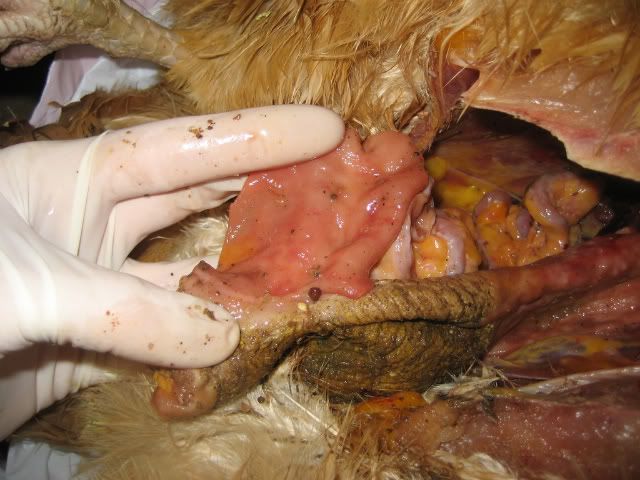
Here is the mesentary, which is the tissue that attaches the intestine loosely together. All the little white dots are very hard and abnormal. All the yellow material is fat in the abdomen. You can see some normal intestine in the picture right near where my index finger is.
I did not submit tissue to the pathology lab, but this looks like some form of intestinal lymphoma with metastasis to the mesentary. Her lungs, heart, liver all looked normal. There was another thickened section another 6-8 inches down the intestine. I don't know if this was a result of lymphoid leukosis, what I have been able to read about that disease makes it sound like it mainly affects younger chickens. If that is true, I guess this could be a "regular" cancer, one not related to an infectious process. Regardless, this was the end of the road for Ivy.

Ivy with her friends in happier days.

Here is a little tour of the chicken digestive tract and her disease, starting with a picture of Ivy in the clinic.
Ivy in her kennel.
Now for the necropsy pictures (graphic)... I made them big so you could see better detail. I started at her beak and cut down through the digestive tract until I found the problem.
Crop, the surface looks healthy and there is no evidence of fungal infection. It was full of greenish/yellow fluid. The red is a postmortem change, it was not like that when I first opened her up.
The glandular stomach (proventriculus) is on the right, the gizzard is on the left. Both were packed full of seeds, grains, little rocks, grass, layer pellet material. Again, the red in the proventriculus is a postmortem change.
This is the intestine just after it left the gizzard. I was packed full of the same material found in the gizzard and proventriculus. The walls are very thin and stretched.
Here is the problem. Her small intestine about 4 inches below the gizzard had very thickened walls and the inside diameter of the intestine was about 3mm. Only liquids could get through. I could hardly get my little scissors through the hole before I cut it open. The whole section in my hand is diseased, the worst area is right above my ring finger in the picture.
Here is the mesentary, which is the tissue that attaches the intestine loosely together. All the little white dots are very hard and abnormal. All the yellow material is fat in the abdomen. You can see some normal intestine in the picture right near where my index finger is.
I did not submit tissue to the pathology lab, but this looks like some form of intestinal lymphoma with metastasis to the mesentary. Her lungs, heart, liver all looked normal. There was another thickened section another 6-8 inches down the intestine. I don't know if this was a result of lymphoid leukosis, what I have been able to read about that disease makes it sound like it mainly affects younger chickens. If that is true, I guess this could be a "regular" cancer, one not related to an infectious process. Regardless, this was the end of the road for Ivy.
Ivy with her friends in happier days.
Last edited:
