- Jun 15, 2008
- 22
- 7
- 24
I ended the battle to help this hen get well. She was suffering too much. A necropsy confirms that. Below are the graphic pics in case it helps someone.
After much reading, I think this hen may have been doomed from shortly after birth. She was given to me by a city hen keeper who said the other hens picked on her too much. Her age was about 7 months. She had been basically hand raised, and never foraged with my other hens, although I never saw them pick on her. She stayed right by the feed tray and ate too much and excercised too little. I think it led to issues with her liver, which eventually failed. I also think the period of wet weather we had, where the feed got wet and the water got too dirty, helped put her over the edge. Although she did have lice, which I treated, and although I treated her for other worms, I don't think any of that caused her troubles. I only gave her 1 treatment for cocci because the vet said the number she saw on the slide (2) could not be significant enough to cause the issues.
Anyway, here's the results. Feedback/observations welcomed:
1.) She had become so bloated, she didn't stand for three days:

2.) Abdomen distended:

3.)

4.) 1st layer removed:

5.)

6.) 2nd layer removed.

7.) Note the fluid filled sac at 3:00.

8.) You should be able to click for a larger image. There are multiple fluid-filled sacs, as well as the fluid in the abdominal cavity.

9.)

10.)

11.)

12.)

13.)

14.) The egg shaped balloon at the bottom is filled with air, nothing more.

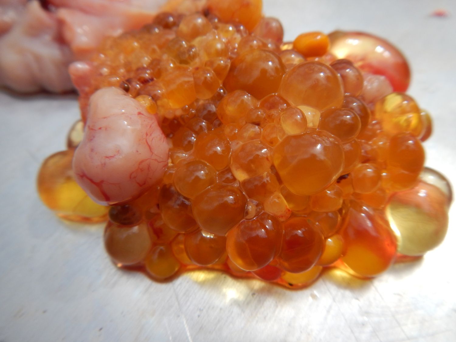
15.) Future eggs. She had not layed since she became ill 5 weeks ago. As I understand it, she had more than she should have had in the same stage of development.

16.)

17.)

18.) This is the amount of fluid that was in her abdomen:

19.)
20.) Heart:

21.) Liver:

22.)

23.)

24.)

25.) Gizzard.

26.)

After much reading, I think this hen may have been doomed from shortly after birth. She was given to me by a city hen keeper who said the other hens picked on her too much. Her age was about 7 months. She had been basically hand raised, and never foraged with my other hens, although I never saw them pick on her. She stayed right by the feed tray and ate too much and excercised too little. I think it led to issues with her liver, which eventually failed. I also think the period of wet weather we had, where the feed got wet and the water got too dirty, helped put her over the edge. Although she did have lice, which I treated, and although I treated her for other worms, I don't think any of that caused her troubles. I only gave her 1 treatment for cocci because the vet said the number she saw on the slide (2) could not be significant enough to cause the issues.
Anyway, here's the results. Feedback/observations welcomed:
1.) She had become so bloated, she didn't stand for three days:
2.) Abdomen distended:
3.)
4.) 1st layer removed:
5.)
6.) 2nd layer removed.
7.) Note the fluid filled sac at 3:00.
8.) You should be able to click for a larger image. There are multiple fluid-filled sacs, as well as the fluid in the abdominal cavity.
9.)
10.)
11.)
12.)
13.)
14.) The egg shaped balloon at the bottom is filled with air, nothing more.
15.) Future eggs. She had not layed since she became ill 5 weeks ago. As I understand it, she had more than she should have had in the same stage of development.
16.)
17.)
18.) This is the amount of fluid that was in her abdomen:
19.)
20.) Heart:
21.) Liver:
22.)
23.)
24.)
25.) Gizzard.
26.)


