This is my documentation of an eggtopsy performed on a late quitting chicken egg.
The back story:
I had 7 eggs under a broody hen. Circumstances arose that required the eggs be moved to an incubator at days 15-16, 20 in a staggered hatch. Two hatched on time, but one was suspected to have quit around day 20. There was veins and faint movement on day 19. By day 21 aircell was unchanged, no drawdown, no movement, skinny veins were seen and an odd patch in the air cell. I put a pinsized safety hole in top of air cell and kept the humidity 65- 70%. Day 22 the opaque patch was smaller and some light presented opposite to air cell, no odor so I went to the morning of Day 23 to be certain the egg had quit before opening.
I performed an eggtopsy in order to determine cause of failure and to educate myself on anatomy and anatomical positioning at late stages of chick incubation. The descriptions of findings are presented above each picture.
Warning- graphic pictures and descriptions.
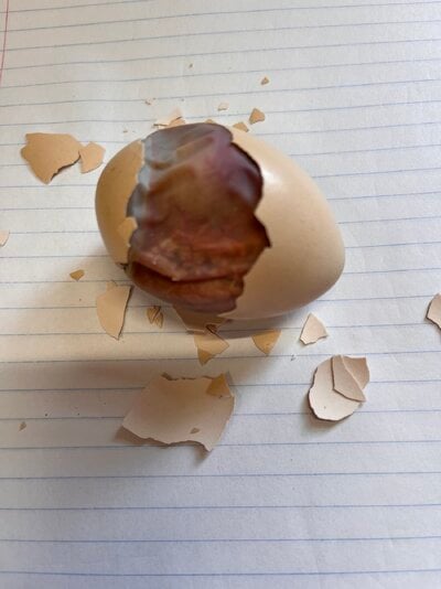
I attempted to remove shell completely using smooth and toothed
forceps, keeping inner membrane intact. The membrane was adhered well to the very top of the shell over the air cell that had the safety hole and so was not removed until later
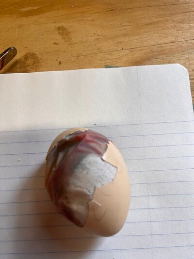
Removing shell from the bottom half of the egg revealed an opaque patch on the membrane that was seen during candling; there were what looked like ‘pores’ on the membrane. The patch was not circumferential and the material was not able to be gently scraped but was thicker than the rubbery membrane. It was likely calcium transfer but was not chalky. The overlying inner shell contained small inward projections that corresponded to the pores on the membrane where the patch was located (zoom in on shell fragment). At the bottom of the egg there is purulent material visible within the intact membrane.


Most of the shell has been removed. The membrane overlying the air cell was breached and yellow/green yolk flowed out. It is difficult to see in the photo but the orientation of the chick looked like it was nearing internal pip into air cell.

Membrane was opened. There was a pungent odor. Immediately I noted caseous material at the bottom of the egg and attached to the bottom of the chick. The feet and legs were intact but the vent, umbilicus and abdomen were less distinguishable in this position from the macerated material. This looked and smelled like tissue in early decay. Green yolk and fluid was present. Note the opaqueness of yolk at the bottom compared to that at the top where paper lines are visible under the latter but not the former.

I flipped the chick and oriented it onto its right side. It looked intact and had an egg tooth formed. Blood vessels and the ruptured yolk sac was visible. The intestines at the umbilicus was visible and quite fragile. This is a female Bielefelder having the chipmunk pattern; Bielefelder is an autosexed breed. I left the chick intact.

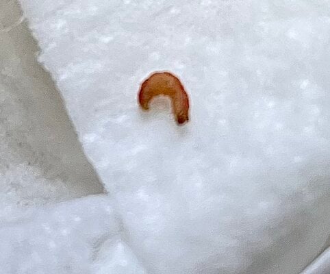
I was not expecting to find any visible parasites but this larvae (presumably) was wriggling within the mass of tissue and fluid. I do not know what it is and no others in my flock have shown signs of parasites.

The eggtopsy taught me a lot about the late stages in chick development, particularly spacial positioning inside the egg.
I do not know the significance or normalcy of the white membrane patch but will look into researching other instances. My suspicion is it is simply calcific transfer from the shell to the membrane, and it had little to do with cause of failure to hatch.
The unexpected larvae is more likely implicated in failure but at what point it in development I cannot know.
The macerated caseous material on the chicks left lower abdomen, vent and membrane, along with foul purulent contents is evidence of dead tissue 48 up to 72 hours duration. Gangrenous gas would have likely formed within the next 12-24 hours risking ruture if not opened.
The back story:
I had 7 eggs under a broody hen. Circumstances arose that required the eggs be moved to an incubator at days 15-16, 20 in a staggered hatch. Two hatched on time, but one was suspected to have quit around day 20. There was veins and faint movement on day 19. By day 21 aircell was unchanged, no drawdown, no movement, skinny veins were seen and an odd patch in the air cell. I put a pinsized safety hole in top of air cell and kept the humidity 65- 70%. Day 22 the opaque patch was smaller and some light presented opposite to air cell, no odor so I went to the morning of Day 23 to be certain the egg had quit before opening.
I performed an eggtopsy in order to determine cause of failure and to educate myself on anatomy and anatomical positioning at late stages of chick incubation. The descriptions of findings are presented above each picture.
Warning- graphic pictures and descriptions.
I attempted to remove shell completely using smooth and toothed
forceps, keeping inner membrane intact. The membrane was adhered well to the very top of the shell over the air cell that had the safety hole and so was not removed until later
Removing shell from the bottom half of the egg revealed an opaque patch on the membrane that was seen during candling; there were what looked like ‘pores’ on the membrane. The patch was not circumferential and the material was not able to be gently scraped but was thicker than the rubbery membrane. It was likely calcium transfer but was not chalky. The overlying inner shell contained small inward projections that corresponded to the pores on the membrane where the patch was located (zoom in on shell fragment). At the bottom of the egg there is purulent material visible within the intact membrane.
Most of the shell has been removed. The membrane overlying the air cell was breached and yellow/green yolk flowed out. It is difficult to see in the photo but the orientation of the chick looked like it was nearing internal pip into air cell.
Membrane was opened. There was a pungent odor. Immediately I noted caseous material at the bottom of the egg and attached to the bottom of the chick. The feet and legs were intact but the vent, umbilicus and abdomen were less distinguishable in this position from the macerated material. This looked and smelled like tissue in early decay. Green yolk and fluid was present. Note the opaqueness of yolk at the bottom compared to that at the top where paper lines are visible under the latter but not the former.
I flipped the chick and oriented it onto its right side. It looked intact and had an egg tooth formed. Blood vessels and the ruptured yolk sac was visible. The intestines at the umbilicus was visible and quite fragile. This is a female Bielefelder having the chipmunk pattern; Bielefelder is an autosexed breed. I left the chick intact.
I was not expecting to find any visible parasites but this larvae (presumably) was wriggling within the mass of tissue and fluid. I do not know what it is and no others in my flock have shown signs of parasites.
The eggtopsy taught me a lot about the late stages in chick development, particularly spacial positioning inside the egg.
I do not know the significance or normalcy of the white membrane patch but will look into researching other instances. My suspicion is it is simply calcific transfer from the shell to the membrane, and it had little to do with cause of failure to hatch.
The unexpected larvae is more likely implicated in failure but at what point it in development I cannot know.
The macerated caseous material on the chicks left lower abdomen, vent and membrane, along with foul purulent contents is evidence of dead tissue 48 up to 72 hours duration. Gangrenous gas would have likely formed within the next 12-24 hours risking ruture if not opened.