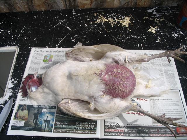
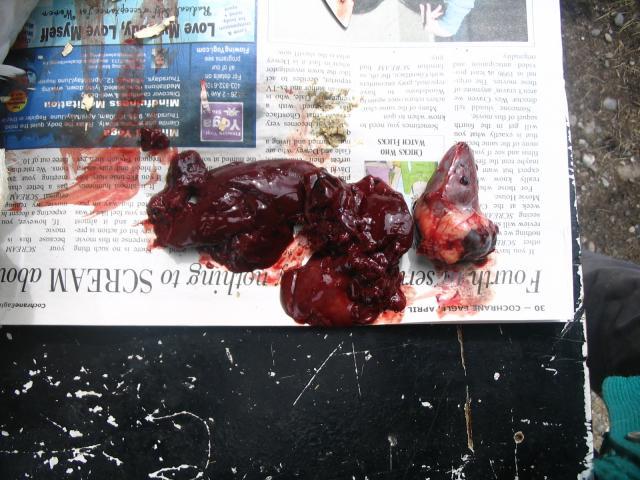
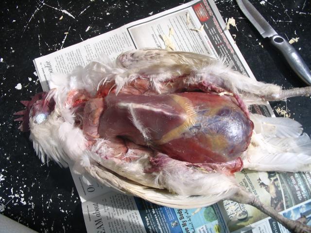
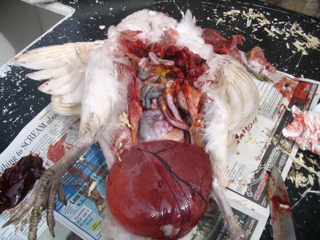
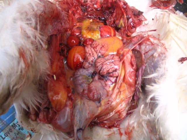
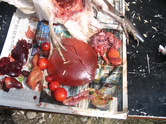
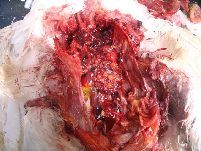
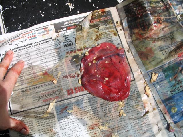
Water Belly or Ovarian Cyst? AND Internal Layer? GRAPHIC PICS

Pics
EXTREMELY GRAPHIC! I have attached autopsy pictures at the end of this post to illustrate her condition. Look or don't look, but I felt, for the sake of informative discussion, it had to be done.

One of my girls developed a hugely swollen and squishy abdomen and lost all the feathers in that area. I researched the condition on here, but couldn't find a description or picture that exactly matched her condition. I did find some posts about draining fluid with a needle or catheter, but was unsure of how or where to insert the needle, as I didn't want to hit anything vital or cause more damage. I was going to start a post about this to see what I could do to help her, but unfortunately, I found her dead yesterday morning. And so I performed my first ever CHICKEN AUTOPSY to find out exactly what and where the mass was. I now know that I could have inserted the needle pretty much anywhere in her lower abdomen without hitting any organs, but the fluid WAS NOT free-floating as I had expected, but was encased in a sack of tissue like a water balloon - I wonder if I might have done more damage by inserting a needle to drain, and thus allowing the fluid to seep into her abdomen from the ruptured sack.

I don't cull my birds and so had no idea what a chicken looked like or is supposed to look like on the inside. This has been an eye-opening experience and I have several questions for you all.

First, as mentioned, the water was not free-floating in the abdomen. It was contained in a veined sack that was attached to the lower right side of the abdomen. When I cut through the abdominal wall, the sack rolled out like a water balloon, exposing the intestines and left oviduct, which were beneath. Smaller water sacks were beneath the main sack, but attached to the same structure. There was a tiny amount of creamy fluid (infection? no smell) within the abdomen, but barely - not as much as I would expect if it had been a massive infection. The sack itself contained clear, odorless fluid.

Crop, liver, spleen, gall bladder, gizzard all appear a normal healthy color - no spots, hard tissues, or off-colors. Crop and gizzard contents as expected for a healthy, feeding chicken. No sour smells and no sign of intestinal parasites. Heart is kind of odd (I think). It seems rather large and appears to bulge on the upper right side.

Ovaries functioning - BUT are they normal? Clusters of dozens of little yolks in various stages of development attached to ovary. Yolks everywhere. Yolks of various sizes passing through left oviduct - no sign of shell formation though. Several loose yolks encased in red tissue (kind of look like cherry tomatoes) of various sizes loose in abdomen. I'm not sure whether this was their original position, or if they rolled from their original location when the mass was released.

So my questions are:
1: Is this truly 'water belly?' I'm wondering because the water was encased in some type of structure and not free floating in the abdomen.
2: Could this tissue sac have been the right oviduct?

I've read that water belly or 'ascites' can be caused by a couple of things, including high altitude and unstable temperatures - puts too much strain on the heart which causes a build up of fluids, which leak from the liver into the abdomen. Large broilers and breeds developed for high egg production are especially susceptible and while there is no cure, the symptoms can be managed by draining the fluid regularly. But is that what this is?

3: Is the above statement accurate?
4: Is the shape of the heart a sign of congestive heart failure?
5: Is the fluid build up associated with the above?
6::Could this be an cyst in the oviduct?
7: If so, what causes this????
8: And what can be done about it?
9: Also: Might she have been an internal layer or is this normal egg development? If she was laying internally, what are the initial symptoms and what can be done?


NOTE: This was not easy for me - I love my girls - but I felt I needed to do it to help me learn to help them. She did receive a proper cremation following the autopsy.


I will never look at my favorite kitchen knife the same way again...
sad.png


WARNING: BELOW ARE EXTREMELY GRAPHIC PICTURES - I appreciate any comments on her condition.


This is what her abdomen looked like. This condition became noticeable rather suddenly - I'd say within a month.
84832_img_0001.jpg


This is a picture of her heart and liver, etc. Note the bulge on the upper part of the heart - congestive heart failure?
84832_img_0008.jpg


The next two images show the fluid-filled mass that was inside her abdomen:

#1: In place (before I cut through the abdominal wall). Note how it fills the entire abdominal cavity.
84832_img_0003.jpg


#2: After I opened the abdominal wall, the mass just rolled out like a water balloon. Note that it is enclosed is some type of tissue (right oviduct?) and attached to the lower right side of the abdomen.
84832_img_0011.jpg


The next several images show egg development.

There were a series of egg yolks of different sizes inside the left oviduct, but also several free-floating yolks throughout her abdomen. Not sure if these may have rolled out of position after I removed the intestines, or if they truly were scattered about like this.
84832_img_0017.jpg


This picture shows the water balloon, the left oviduct (upper right) with yolk enclosed, and several free-floating cherry red yolks that were found throughout the abdomen (in places where I wouldn't think they could reach the oviduct)
84832_img_0021.jpg


This, I'm assuming, is a mass of yolks developing in her ovaries. Is this normal production? Until recently, this hen laid an egg every day - sometimes twice a day.
84832_img_0022.jpg


This image is the mass after it was drained. The fluid drained was clear, odorless and a little thicker than water.
84832_img_0024.jpg


Thanks for any feedback.
Hope you are still on BYC because you did one hell of a job here. Very very informative. did you ever find out more info on this condition ? I did have a girl the vet autopsied she definitely had many yolks peritonitis that took her.
 

New posts New threads Active threads

Back
Top Bottom