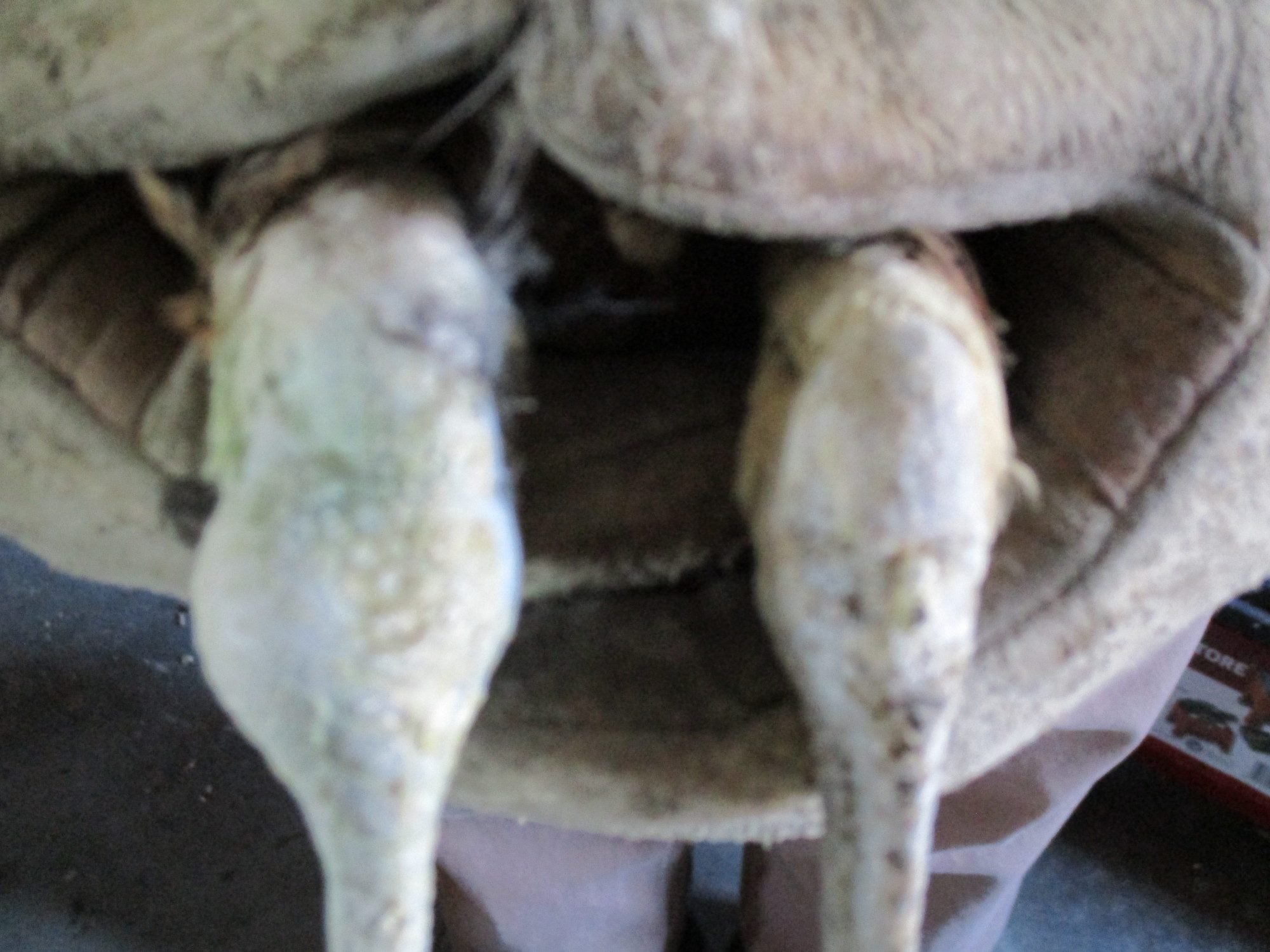
Follow along with the video below to see how to install our site as a web app on your home screen.
Note: This feature may not be available in some browsers.
Quote:
Welllll ---
It's really hard for me to interpret from pictures, without seeing, feeling, manipulating and smelling it myself, but I'll stick my toe in the water (since everybody has popcorn, after all).
The first pictures look like the lesion is below the joint, not in it. The picture where the joint is disarticulated looks like the articular cartilage was not inflammed, and there is a clear glistening of what I assume is normal joint fluid. The surrounding area does look a little thickened, which I assume is inflammation moving upwards from the lesion and surrounding the joint, but hasn't invaded into the protective joint capsule yet. It appears that the swelling started distinctly below the hock joint (the knee is the next one up the leg, and points towards the head when flexed, whereas the hock, the equivalent to our ankle, points towards the tail when flexed), with the greatest amount of inflammation on the back of the leg, with progressively thinning areas of swelling wrapping mostly around the outside, but somewhat also around the inside of the leg, the extending upwards toward the joint as well.
The description of the center of the swelling being hard and crunchy means that either bony tissue is involved, or there is some kind of crystal formation, or mineralization of soft tissue, or some combination of those. Other things that I would consider are the lack of a distinct tubular bone structure, very inflammed/thickened connective tissue outside the bone (probably either the (normally) thin periosteum that surrounds the outside of the mineralized bone, or the ends of tendons that were attached to the bone in that location), the level of pain the bird showed (laying down instead of limping), and the sudden change in the birds condition from being disabled to being really sick. That bone, focally, was really compromised and desperately remodeling with a huge amount of disorganized bone tissue, instead of healing with a strong, functional bony structure. That kind of bone growth is kind of like rapidly building something with styrofoam instead of slowly creating steel. The repair takes up more space and isn't nearly as strong. That rarely happens with a fracture. I suppose it could if the animal was forced to use the leg with an unstable fracture, where the broken bone ends aren't held together and they rub against each other with each step. But typically you would have a bird that suddenly went down, before the extreme bony swelling had a chance to develop, and you would first see soft tissue swelling from the trauma and maybe be able to detect the instability. A green stick fracture shouldn't have that level of instability, so the bony callous that it develops shouldn't be that exuberant, and the inflammation shouldn't be so extreme. And there should still be an obvious strong bony "pipe" that the new mineralization is adhering to and trying to repair. So to my eyes, this doesn't look like a typical green stick fracture, although I can't be sure from just pictures.
The two things that first come to mind that would cause this level of disorganized bony proliferation are a bone infection (bacterial or fungal are most common), or bone cancer. Depending on where you live, fungal infections may be common, can seat anywhere on a bone, and will look exactly like this. Bacterial infections can get into bone through injury and a tiny penetrating wound that might never be seen, or can land focally on a bone after traveling through the blood. An injury that would cause a green stick fracture plus a tiny penetrating wound leading to bone infection would be a double whammy. These types of infections don't look like typical abscesses, don't form pus, and rarely smell bad. This bird is a little young for cancer, but realistically, they have pediatric oncology wards for a reason. Youngsters get cancer too, unfortunately, and bone cancer typically causes first lytic resorption of bone tissue, then desperate disorganized attempts at repairing of the bone, ending in severe focal pain, then obvious illness. So either infection or cancer could look exactly like this. Gout could potentially also cause this, but the location is weird, the progression isn't right, and the response would be extreme for just a few uric acid crystals. This level of gout typically would take a very long time to develop, and should not be painful enough outside of a joint to create these symptoms. I could see this level of inflammation if gout settled in a tendon, but that shouldn't trigger this kind of bony remodeling. A "slipped tendon" causes some obvious abnormal angulation of the leg at the hock joint, and may cause some local soft tissue inflammation and swelling, but would have no reason to cause bony remodeling to this extent. Something had to be destroying bony tissue to justify this kind of a response.
Or at least that's my best guess, based just on pictures and a few symptoms. I could be wrong.
If the dissected area was saved, you could send it in for a biopsy. That would likely give you a more definitively answer.
Is he valuable? If so, maybe an X-ray?
-Kathy
It is a hi% Spaulding Black Shoulder, cock I think... $175 ish in healthy condition. I have a few more but spending a bunch of money on this one is not really appealing to me. BTDT... I will call and see how much an X-ray is. If it is a hen I will be more inclined to get that X-ray. Gerald has a full brother to this one and he really likes his cock.