The gram stain is an indispensable test used frequently in microbiology, to help determine if said sample contains bacteria, and divide the bacteria into two classes, Gram-positive, and gram-negative. The test is very simple, can be performed by almost anyone, and requires very few supplies. The point of this thread is to provide insight to owners looking to perform this test on their waterfowl to ultimately aid in the diagnosis, and treatment of the problem, and rule out other possible conditions.
To start, a general understanding of common bacteria in waterfowl needs to be established, and how to read the results of a gram stain. When properly done, gram-positive bacteria should appear a dark purple on a gram stain, while gram-negative bacteria often appear a pink/light purple. In birds, commonly seen gram-positive bacteria include staphylococcus, streptococcus, clostridium, and bacillus. Common gram-negative bacteria seen include Escherichia coli, Pseudomonas, Klebsiella, Pasteurella, among many more. While properly performed gram stain allows you to differentiate gram-positive bacteria from gram-negative bacteria, in some cases, basing on the bacteria morphology, a presumptive direct identification can be accomplished by examining the morphology of the bacteria on the slide. For example, if one were to stain a slide, and see mostly gram-positive coccus-shaped bacteria, forming grape clusters, odds are the bacteria is of the staphylococcus family. Members of the clostridium family, often have a drumstick-like appearance. To differentiate members of the staphylococcus genus, the bacteria would need to be grown on agar, and other microbiological tests, like blood agar, coagulase, and novobiocin resistance would need to be determined.

https://commons.wikimedia.org/wiki/File:Bacterial_morphology_diagram-hu.svg
As shown in the graph above, bacteria can appear in several shapes and forms on a gram stain. One must take the time to understand the different types of shapes bacteria can come in, so the stain can be properly read. The most common bacteria shapes that will be noted, will be cocci (spherical-shaped), and bacilli (hot dog-shaped) rods.
A gram stain can be performed on several sites, including the mouth, tumors, tissue, organs, and fecal samples which is the one I will discuss here. Whenever collecting a sample, care must be taken not to contaminate the sample as incorrect readings may occur. To take a fecal sample, either wait till the bird defecates, or rub a blunt wooden pick around the cloaca to induce defecation. Once a sample has been collected, dispense the sample into a sterilized container until the staining can be done.
To stain a sample, one must have several dyes to do the gram stain, including a 1000x microscope, inoculating loop, coverslips, and a fixator such as a bunsen burner, are more preferred methanol.
To prepare a stain for fecal matter, grab some of the fecal matter with a small pick, add a few drops of saline, then transfer one drop of the liquid using a sterilized loop onto a slide marked with the patient's name, and a circle on the underside of the slide so when viewing on the microscope you know where to position the scope.
Spread the drop thinly on the slide using an inoculating loop so faster drying time is achieved, and so the bacteria is thinned out when viewing it on the slide. Once the sample has dried thoroughly, the bacteria need to fixed to the slide when applying the dyes mentioned above, so the bacteria doesn't wash away in the process. Two ways can be done to fix the bacteria to the slide, one is done by passing the slide through an active flame several times until the slide is warm to the touch. An alternative way, which now is the preferred method as it increases cytoplasmic clarity in the sample, is to add a few drops of methanol to the slide, and allow to air dry for a few minutes.

Now that you have a properly collected sample, the has been fixed to the slide you may begin the staining procedure. Set the slide on a wire rack over a container, and add a few drops of gentian violet to the sample. Wait one minute, and gently wash off the violet with distilled water. This step has dyed all bacteria on the sample purple. The next step includes adding a few drops of grams iodine to the sample to improve better bonding of the dye with the cell membrane. As with the violet, wait one minute, then gently wash off, not directing the stream towards the bacteria, but rather letting the water flow down. After the iodine has been washed off you will begin the decolorization process which washes out the violet stain of the gram-negative bacteria, but not the gram-positive bacteria as gram-negative bacteria have thin cell walls. This step is very prone to errors, so if it's your first time, I would suggest acquiring bacteria samples of a known class so you can practice making sure it is staining correctly. To decolorize, hold the slide upright and apply a few drops of the decolorizer until the drops dripping off the slide are clear. This process should take only ten seconds. Wash the slide after done decolorizing. At this point, the only stained bacteria are the gram-positive bacteria, so safranin is added to the slide for one minute to stain the gram-negative bacteria.
Wash off the remaining safranin, and set the slide upright on a rack, and allow to air dry. Once, the slide is dry, set the slide on the microscope plate with the bacteria stained slide facing upright. I generally, start off with a 50x lens, and assess the arrangement of the bacteria, and move closer to 100x to assess proper color.
Note the arrangement of the cells. Are they in grape clusters what's the density, are they forming chains, and what color is the sample?
With fecal samples, bacteria on the slide should mostly consist of gram-positive bacilli. The cells should be fairly spread out. If you see a fairly dense slide of bacteria and are also seeing an overabundance of gram-negative (pink bacteria). There is likely something at play, and further samples should be done, so a culture and sensitivity test can be performed to further identify the bacteria, and identity its sensitivity to certain antibiotics.

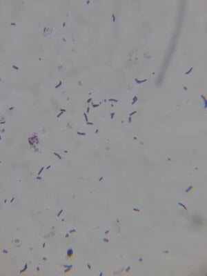
: The picture showed above was a fecal sample taken from a healthy duck. Note the gram-positive bacilli rods, and how the bacteria spread out well.

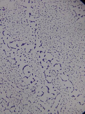
: The picture showed above is a gram stain of tissue infected with staphylococcus. Note the grape clusters and the small cocci like cell shape, when zoomed in closer.

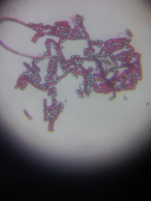
: The picture showed above was an oral sample taken from a canine, consisting of gram-negative bacteria. Through culture, and other microbiological identification tests, the bacteria were later identified as Moraxella. sp.
I have found this source to be quite helpful in learning about gram stains :
http://avianmedicine.net/wp-content/uploads/2013/03/gramstain2.pdf
To start, a general understanding of common bacteria in waterfowl needs to be established, and how to read the results of a gram stain. When properly done, gram-positive bacteria should appear a dark purple on a gram stain, while gram-negative bacteria often appear a pink/light purple. In birds, commonly seen gram-positive bacteria include staphylococcus, streptococcus, clostridium, and bacillus. Common gram-negative bacteria seen include Escherichia coli, Pseudomonas, Klebsiella, Pasteurella, among many more. While properly performed gram stain allows you to differentiate gram-positive bacteria from gram-negative bacteria, in some cases, basing on the bacteria morphology, a presumptive direct identification can be accomplished by examining the morphology of the bacteria on the slide. For example, if one were to stain a slide, and see mostly gram-positive coccus-shaped bacteria, forming grape clusters, odds are the bacteria is of the staphylococcus family. Members of the clostridium family, often have a drumstick-like appearance. To differentiate members of the staphylococcus genus, the bacteria would need to be grown on agar, and other microbiological tests, like blood agar, coagulase, and novobiocin resistance would need to be determined.
https://commons.wikimedia.org/wiki/File:Bacterial_morphology_diagram-hu.svg
As shown in the graph above, bacteria can appear in several shapes and forms on a gram stain. One must take the time to understand the different types of shapes bacteria can come in, so the stain can be properly read. The most common bacteria shapes that will be noted, will be cocci (spherical-shaped), and bacilli (hot dog-shaped) rods.
A gram stain can be performed on several sites, including the mouth, tumors, tissue, organs, and fecal samples which is the one I will discuss here. Whenever collecting a sample, care must be taken not to contaminate the sample as incorrect readings may occur. To take a fecal sample, either wait till the bird defecates, or rub a blunt wooden pick around the cloaca to induce defecation. Once a sample has been collected, dispense the sample into a sterilized container until the staining can be done.
To stain a sample, one must have several dyes to do the gram stain, including a 1000x microscope, inoculating loop, coverslips, and a fixator such as a bunsen burner, are more preferred methanol.
To prepare a stain for fecal matter, grab some of the fecal matter with a small pick, add a few drops of saline, then transfer one drop of the liquid using a sterilized loop onto a slide marked with the patient's name, and a circle on the underside of the slide so when viewing on the microscope you know where to position the scope.
Spread the drop thinly on the slide using an inoculating loop so faster drying time is achieved, and so the bacteria is thinned out when viewing it on the slide. Once the sample has dried thoroughly, the bacteria need to fixed to the slide when applying the dyes mentioned above, so the bacteria doesn't wash away in the process. Two ways can be done to fix the bacteria to the slide, one is done by passing the slide through an active flame several times until the slide is warm to the touch. An alternative way, which now is the preferred method as it increases cytoplasmic clarity in the sample, is to add a few drops of methanol to the slide, and allow to air dry for a few minutes.
Now that you have a properly collected sample, the has been fixed to the slide you may begin the staining procedure. Set the slide on a wire rack over a container, and add a few drops of gentian violet to the sample. Wait one minute, and gently wash off the violet with distilled water. This step has dyed all bacteria on the sample purple. The next step includes adding a few drops of grams iodine to the sample to improve better bonding of the dye with the cell membrane. As with the violet, wait one minute, then gently wash off, not directing the stream towards the bacteria, but rather letting the water flow down. After the iodine has been washed off you will begin the decolorization process which washes out the violet stain of the gram-negative bacteria, but not the gram-positive bacteria as gram-negative bacteria have thin cell walls. This step is very prone to errors, so if it's your first time, I would suggest acquiring bacteria samples of a known class so you can practice making sure it is staining correctly. To decolorize, hold the slide upright and apply a few drops of the decolorizer until the drops dripping off the slide are clear. This process should take only ten seconds. Wash the slide after done decolorizing. At this point, the only stained bacteria are the gram-positive bacteria, so safranin is added to the slide for one minute to stain the gram-negative bacteria.
Wash off the remaining safranin, and set the slide upright on a rack, and allow to air dry. Once, the slide is dry, set the slide on the microscope plate with the bacteria stained slide facing upright. I generally, start off with a 50x lens, and assess the arrangement of the bacteria, and move closer to 100x to assess proper color.
Note the arrangement of the cells. Are they in grape clusters what's the density, are they forming chains, and what color is the sample?
With fecal samples, bacteria on the slide should mostly consist of gram-positive bacilli. The cells should be fairly spread out. If you see a fairly dense slide of bacteria and are also seeing an overabundance of gram-negative (pink bacteria). There is likely something at play, and further samples should be done, so a culture and sensitivity test can be performed to further identify the bacteria, and identity its sensitivity to certain antibiotics.

: The picture showed above was a fecal sample taken from a healthy duck. Note the gram-positive bacilli rods, and how the bacteria spread out well.

: The picture showed above is a gram stain of tissue infected with staphylococcus. Note the grape clusters and the small cocci like cell shape, when zoomed in closer.

: The picture showed above was an oral sample taken from a canine, consisting of gram-negative bacteria. Through culture, and other microbiological identification tests, the bacteria were later identified as Moraxella. sp.
I have found this source to be quite helpful in learning about gram stains :
http://avianmedicine.net/wp-content/uploads/2013/03/gramstain2.pdf


