The Anatomy and Physiology of the Chicken
When you own a chicken, it is very important to understand the anatomy and physiology of your bird. Anatomy is the science of the structure of animals. Physiology is the science that deals with the functions of the living organism and its parts. Both of these works in tandem with each other to keep your chicken -and all other living organisms- alive and well. When something goes wrong in any part of either the anatomy or the physiology, complications arise -often resulting in the illness or death of the organism.
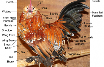
The easiest anatomy of the chicken to assess is the external anatomy. This is the plumage and various appendages that can be seen without any further investigation necessary. This includes the plumage, legs, beak, comb, wattles, eyes, toes, tongue, mouth and skin.
Diagram 1 shows the external anatomy of a mature rooster

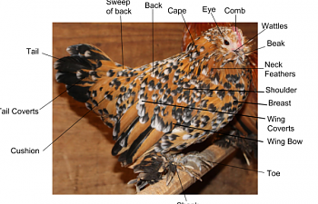
Diagram 2 shows the external anatomy of a mature hen
Not only is it important to recognize and know the positions of certain anatomical features, but it is even more important to understand their functions.
Skin, feathers and the beak all serve to protect the bird from external harm. Feathers are dense and specifically shaped to protect the skin from cuts and abrasions as the bird lays down, is in crowded areas or otherwise does the things that chickens do. The beak closes water and airtight to prevent particles from enter the birds trachea as they breathe. When the bird opens its beak, this function is temporarily lost. The skin is the largest organ on any living animal and, as with all species of animal, the skin serves as the first triage for harmful pathogens. When intact, pathogens cannot enter the body through the tissue and good bacteria on the surface of the skin helps to kill off most pathogens. In birds, the skin also acts as a host for feather follicles and an anchor for feather growth.
The comb and wattles serve to regulate the internal temperature of the chicken through the cooling of blood. When a chicken is hot blood rushes into the comb -causing it to turn a brighter red- and the subsequent exposure to cooler air cools the blood in the tiny capillaries that run throughout the comb and wattles. The cooled blood then returns to the inside of the chicken, where it continues to circulate throughout the body. When a chicken is cold its comb becomes paler as the body draws blood from the extremities towards the internal organs to maintain function of critical organs.
The eyes serve as vehicles for vision. The eyelids -of which chickens have three- serve as a level of protection to help prevent harmful pathogens from entering the bloodstream. The outer eyelids clear the eye of debris, while the inner eyelid serves as the 'blinking' mechanism and helps to keep eyes from drying out.
In addition to the aforementioned anatomy, the anatomy of a chicken's wing also has several parts that contribute to the whole. While chickens are generally flightless creatures, they still retain the wing feather anatomy of other birds of flight -just in smaller versions.
Diagram 3 shows the anatomy of the wing plumage
There are seven main parts to the wing, as can be observed above. Each part has a purpose. More information about other feathers on a chicken can be found here.
Primaries are the longest feathers in the wing. They are the outer feathers of the dominant flight feathers and are the strongest of the flight feathers. The primaries grow outwards from the end of each wing.
Secondaries are positioned behind the primaries and are the inner flight feathers. They grow out form the 'forearm' area of the wing. When it comes to soaring and flapping, the secondaries are the feathers that provide the lift.
Primary and secondary coverts aid in streamlining the wing and providing insulation. The marginal Coverts and Alula serve the same function. The Scapulars also known as the tertiary feathers are the innermost flight feathers and are not as important to flight as the primaries and secondaries.
The next page will discuss the digestive system.
The Digestive System
The Digestive System
The digestive system of the chicken starts at the mouth and ends at the cloaca, with multiple intermediate organs. Each organ in the digestive system plays a critical role and though a chicken may be able to survive with the damage or loss of certain organs, the resulting effects of such damage or loss could be fatal.
Among chicken illnesses, the most common ailments tend to take root in the digestive system of the bird. Included wuold be cocci, impacted crop, sour crop, impacted gizzard, intestinal parasites, and blockages of the intestine.
Seeing as a chicken's energy comes from food and water and the digestion of nutrients transported in those forms, it is imperative to understand the structure and functions of the entire digestive system. Knowing what is normal in regards to the digestive system should help cultivate your understanding of the nutritional requirements of your chicken as well as when something is wrong with the digestive tract -hopefully before it is too late.
Diagram 4 shows the illustrated digestive system of a chicken
Whether a chicken forages for their food or is fed a complete diet, the bird still has to ingest and digest the feed. This process takes a while and the food travels through many organs, changes shape and texture and is utilized differently in different sections of the digestive tract.
The digestive tract begins with the beak, but digestion begins with the mouth. Although chickens pick up feed with their beaks, the feed enters the digestive system through the mouth. The mouth is made up of glands and enzymes. The glands in the mouth secrete saliva, which contains the digestive enzyme amylase that starts to digest the feed upon contact. The tongue's function is to push the feed to the back of the mouth so that it can be swallowed.
The feed then enters the esophagus -a flexible tube which connects the mouth with the rest of the digestive tract. The esophagus is long and is present in two transitional areas -from the mouth to the crop, and from the crop to the proventriculus. The function of the esophagus is to transport food between organs in the first half of the digestive system.
Next, the feed reaches the crop, where it is stored for up to 12 hours. The crop is located on the upper right side of the breast of the chicken. The crop can expand to store copious amounts of water and food. Little to no digestion takes place in the crop.
From the crop, the feed travels again through the esophagus into the proventriculus, the glandular stomach where digestion begins. Hydrochloric acid and the digestive enzyme Pepsin are added to the feed in the proventriculus and further breakdown of the feed takes place.
The feed and digestive juices then pass from the proventriculus to the gizzard, which is the mechanical stomach. Here, the food is crushed, mixed and mashed. The gizzard is made up of two sets of strong muscles. These muscles serve the same function that teeth serve us and other mammals who have them. Stored in the gizzard are small insoluble stones, or grit, that aid in the mechanical breakdown of feed.
The now mushy feed passes from the gizzard into the small intestine. The small intestine's function is to absorb the large majority of the nutrients that are useful to the bird. Nutrients such as calcium, lipids, carbohydrates, sugars, potassium, sodium, vitamins and minerals diffuse into the surrounding capillaries. Some water absorption also takes place in the small intestine. The small intestine is made up of the duodenum, and the lower small intestine. The duodenum receives digestive enzymes and bicarbonate from the pancreas and bile from the gall bladder. Protein digestion is possible with the digestive enzymes produced by the pancreas. The pancreas also serves to regulate the bird's blood sugar by the secretion of insulin and glycogen which raise and lower blood sugar accordingly. Bile is important to the digestion of lipids and the absorption of fat soluble vitamins A, D, E and K. The remainder of the digestion occurs primarily in the duodenum. The remaining nutrients are absorbed mostly in the jejunum and ileum. Remaining nutrients and their juices are then stored in the ceca, to be deposited later.
The large intestine is the next part of the digestive tract and has little to nothing to do with nutrient absorption. By this point, almost all of the nutrients are absorbed. The large intestine's role is to reabsorb the large amounts of water in the fecal matter, drying up the fecal matter and working to prevent dehydration in the chicken. Urates join the fecal matter in the Cloaca and both urates and fecal matter are deposited at once and removed from the digestive tract.
A comprehensive video, produced and owned by Nutrena, of the chicken's digestive tract is posted below for your learning convenience.
The next page will discuss the Respiratory System.
The Respiratory System
The Respiratory System
In all animals, the respiratory system is responsible for the absorption of oxygen, the release of carbon dioxide, the release of heat, detoxification of certain chemicals, the rapid adjustment of the acid-base balance and vocalization.
Although avian and mammal respiratory systems are very similar in their functions, their anatomy is quite drastically different. Just as with mammals, birds also have two symmetrical lungs that are connected to a trachea. Mammalian lungs contain bronchi, which lead to aveoli. The aveoli in mammalian lungs have only one opening -meaning they don't go all the way through the lung. The avian lung has parabronchi which are continuous tubes that allow for air to pass through the lung in one direction. These parabronchi are laced with capillaries. These capillaries are where the gas exchange occurs.
Diagram 5 shows the illustrated respiratory system of the chicken
The avian respiratory tract begins with the Glottis, which closes when feed is passed down the throat to prevent the inhalation of feed. The glottis connects to the trachea, which is made up of cartilaginous rings to prevents its collapse from the negative pressure caused by inhalation.
The syrinx is the vocal organ of birds. Vocalizations are made by air pressure on a sound valve and modified by muscle tension. The avian lungs are relatively small and they do not expand. The lungs are firmly attached to the rib cage. Birds have an incomplete diaphragm. Instead of being able to expand and deflate lungs, birds have air sacs, which inflate and deflate accordingly. In birds, there are nine such air sacs.
On first inhalation, air flows in from the trachea and bronchi, primarily into the caudal thoracic air sacs.
On exhalation, air moves from the caudal thoracic air sacs into the lungs.
With the second inhalation, air moves from the lungs into the cranial thoracic air sacs.
With the second exhalations, air moves from the cranial thoracic air sacs back into the trachea and out.
The next page will discuss the skeletal system.
The Skeletal System
The Skeletal System
Besides structural support, the skeletal system is responsible for respiration and calcium transport. A chicken's skeletal system is both lightweight and compact, yet strong. The tail and neck vertebrae can move, while the vertebrae in the body are fused together in order to support the wings. Birds have special types of bones: the pneumatic and medullary bones.
Diagram 6 shows a labelled diagram of the chicken skeletal system
The pneumatic bones are important for respiration. They are hollow bones connected to the respiratory system and are important for the chickens to breathe. Examples of some pneumatic bones are the skull, humerus, clavicle, keel, pelvic girdle and lumbar and sacral vertebrae.
The medullary bones are an important source of calcium for a laying hen. Examples include the tibia, femur, pubic bones, ribs, ulna, toes and scapula.
Several key adaptations have been made to the skeleton of the chicken to allow it to be a functional bird. Although chickens arguably can't fly, their skeleton is built for this.
The thoracic vertebrae are fused to support the wings. The skull is smaller because a larger skull is difficult to fly with. The sternum (keel) has a large surface area for the attachment of the main flight muscles. The tail is a short section of fused bones called a pygostyle. The ribs include the ucinate process. These are overlying flaps that connect each rib to give the rib cage support and prevents its collapse during flight.
Just as with humans, the wing is comprised of the radius and ulna, extending from the joint (the elbow) to the metacarpus (the wrist) and from there to the phalanges (the fingers).
The leg is also very similar to that of the leg of a human. The first -and strongest- bone in the leg is the femur (thigh), followed at the joint (kneecap) by the fibula and tibia, which meet another joint (ankle) leading out to metatarsus which meets the ball of the foot and spreads out into the toes.
The next page will discuss the muscle system.
The Muscle System
The Muscle System
A chickens muscle system is comprised of 3 types of muscles. These are the smooth muscles, cardiac muscles, and skeletal muscles. The smooth muscles are found in the blood vessels, intestines and gizzard. The cardiac muscles comprise the muscles of the heart. The skeletal muscles are the muscles that shape the bird and are responsible for voluntary movement. The skeletal muscles are the edible muscles on a carcass.
Diagram 7 shows the muscle structure of a chicken
Muscles are structured from special muscle cells in the form of fibres that have the ability to contract or shorten. When they relax the muscle lengthens.
There are three main types of muscles found in any animal body. These are:
Muscle contractions are the result of a stimulus passing into the muscle fibers. This stimulus causes the myofibril segments to shorten because actin filaments slide across the myosin filaments. Much of the energy taken from digested feed is used in muscle contractions, particularly calcium.
There are white and red types of skeletal muscle fibers found in birds and all muscles have some white fibers and some red fibers. However, the proportion varies and some muscles are predominantly white and others predominantly red or dark. White fibers lack a compound called myoglobin, but store more glycogen and have a fast contraction of short duration. They have little staying power. The breast muscles of fowls, the muscles of flight, are predominantly white fibers and fowls have very poor flying ability. They fly very short distances with a very rapid wing movement. Red fibers have myoglobin and other cellular structures for continuous production of energy for contraction. These fibers have a slow contraction of long duration. The flight muscles of flying birds consist mainly of red fibers.
The next page will discuss the reproductive system.
Diagram 7 shows the muscle structure of a chicken
Muscles are structured from special muscle cells in the form of fibres that have the ability to contract or shorten. When they relax the muscle lengthens.
There are three main types of muscles found in any animal body. These are:
- Involuntary muscles found in the walls of the alimentary canal, blood vessels, air passages and other tubular structures. These muscles are beyond the control of the will and are called involuntary muscles. The fibers of these muscles do not carry transverse striation or stripes and are therefore said to be ‘unstriped’ or ‘unstriated’.
- Cardiac muscle of the heart. This too is involuntary muscle but is striated and is structured differently to other muscle. It is nucleated, contains many Purkinje Fibers and forms a syncytium with many nuclei but no differentiation of the protoplasm into cells.
- The striated or striped, voluntary muscles of the body that move the various parts of the skeleton or appendages. These consist of very minute thread-like muscle fibers in bundles enclosed by sheaths of fibrous tissue.
Muscle contractions are the result of a stimulus passing into the muscle fibers. This stimulus causes the myofibril segments to shorten because actin filaments slide across the myosin filaments. Much of the energy taken from digested feed is used in muscle contractions, particularly calcium.
There are white and red types of skeletal muscle fibers found in birds and all muscles have some white fibers and some red fibers. However, the proportion varies and some muscles are predominantly white and others predominantly red or dark. White fibers lack a compound called myoglobin, but store more glycogen and have a fast contraction of short duration. They have little staying power. The breast muscles of fowls, the muscles of flight, are predominantly white fibers and fowls have very poor flying ability. They fly very short distances with a very rapid wing movement. Red fibers have myoglobin and other cellular structures for continuous production of energy for contraction. These fibers have a slow contraction of long duration. The flight muscles of flying birds consist mainly of red fibers.
The next page will discuss the reproductive system.
The Reproductive System
The Reproductive System
The avian reproductive system is nothing like that of mammals. The reproductive system of aves has evolved to cater to the specific needs and challanges of being a bird. One of the biggest problems of being a bird is that you're everyone's dinner guest. Besides the specific anatomy of the reproductive system itself, birds have adapted a characteristic of reproduction that is very unlike most mammals. Most mammals will have few offspring and will spend copious amounts of time devoted to raising them. Birds, with a few exceptions, will do the opposite -choosing instead to raise large quantities of offspring, devote minimal care, if any, and then leave them to figure things out on their own.
The ovary is a cluster of devoloping yolks, called ova (singular ovum), located midway between the neck and tail of the hen. When a female chick hatches her ovary is fully formed albeit very small. The ovary contains 13,000-14,000 ova that only mature into yolks with the addition of a special fluid called 'yolk fluid'. Each ovum starts out surrounded by a membrane called the vitteline membrane. Over time, yolk is added as the ovum develops. When a female chick hatches she is equipped with all of the eggs that she will ever have.
Ovulation is the release of a mature ovum from the ovary. The ovum is released into the oviduct. The ovum, enclosed in a sac, ruptures along the stigma. The release of the ovum occurs 30-75 minutes after the previous egg is laid.
Diagram 8 shows the female reproductive system
The oviduct is a convoluted tube about 25-27 inches long, which is divided into 5 major sections. These sections are the infundibulum, magnum, isthmus, shell gland/uterus, and the vagina.
The first part of the oviduct, the infundibulum, is 3-4 inches long and it engulfs the ovum that is released from the ovary. The ovum then remains in the infundibulum for 15-18 minutes. The infundibulum is the area in which fertilization can take place.
The next section of the oviduct is the magnum which is 13 inches long. The magnum is the largest section of the oviduct. The ovum remains in the magnum for 3 hours. It is here that the albumen is added.
The third section of the oviduct is the isthmus which is 4 inches long. The egg remains here 75 minutes. The isthmus is slightly constricted. The isthmus is where the inner and outer shell membranes are added.
The next section of the oviduct is the shell gland, or uterus. The shell gland is 4-5 inches long and the egg remains here for over 20 hours. As the name implies, the shell is developed here. The hen sources 47% of her calcium from her bones and her diet supplies the rest. The shell gland is also where the pigment is applied.
The last section of the oviduct is the vagina which is 4-5 inches long and does not play a part in egg formation. The vagina's function is to push the egg out of the body. There are glands located in the vagina where sperm is stored. It is these glands which allow a hen to store sperm for extended periods of time. The bloom is added to the egg in the vagina.
Diagram 9 shows the reproductive system of the male chicken alongside the reproductive system of the female chicken
The male reproductive system and how it functions is almost entirely the same as with mammals, but does a few key differences.
Sperm is produced in the seminiferous tubules which are located in the testes. Sperm production occurs best at slightly cooler temperatures. Because of this, spermatogenesis may occur primarily at night when the body temperature of the bird is lower. Sperm are stored at the terminal end of the vas deferens.
Lying cranioventral to the first kidney lobe are the paired testes. The vas deferens emerges medially and passes caudally to the cloaca where it has a common opening with the ureter in the Urodeum. The terminal vas deferens is swollen as a storage organ: the seminal glomus.
Sperm formation is temperature sensitive. Male birds tend to have relatively low extragonadal sperm reserves and sperm are ejaculated soon after production in the testes.
The next page will discuss the circulatory system.
The female reproductive system
The reproductive system of the female chicken is divided into two main parts: the ovaries and the oviduct. In hens, the left ovary is the only functional ovary. Thus, it is the only ovary secreting hormones and, if damaged, female hormone balance can be thrown off track. The right ovary, however, is present in embryos but regresses during devlopment and serves no purpose in adult hens. The ovary is a cluster of devoloping yolks, called ova (singular ovum), located midway between the neck and tail of the hen. When a female chick hatches her ovary is fully formed albeit very small. The ovary contains 13,000-14,000 ova that only mature into yolks with the addition of a special fluid called 'yolk fluid'. Each ovum starts out surrounded by a membrane called the vitteline membrane. Over time, yolk is added as the ovum develops. When a female chick hatches she is equipped with all of the eggs that she will ever have.
Ovulation is the release of a mature ovum from the ovary. The ovum is released into the oviduct. The ovum, enclosed in a sac, ruptures along the stigma. The release of the ovum occurs 30-75 minutes after the previous egg is laid.
Diagram 8 shows the female reproductive system
The oviduct is a convoluted tube about 25-27 inches long, which is divided into 5 major sections. These sections are the infundibulum, magnum, isthmus, shell gland/uterus, and the vagina.
The first part of the oviduct, the infundibulum, is 3-4 inches long and it engulfs the ovum that is released from the ovary. The ovum then remains in the infundibulum for 15-18 minutes. The infundibulum is the area in which fertilization can take place.
The next section of the oviduct is the magnum which is 13 inches long. The magnum is the largest section of the oviduct. The ovum remains in the magnum for 3 hours. It is here that the albumen is added.
The third section of the oviduct is the isthmus which is 4 inches long. The egg remains here 75 minutes. The isthmus is slightly constricted. The isthmus is where the inner and outer shell membranes are added.
The next section of the oviduct is the shell gland, or uterus. The shell gland is 4-5 inches long and the egg remains here for over 20 hours. As the name implies, the shell is developed here. The hen sources 47% of her calcium from her bones and her diet supplies the rest. The shell gland is also where the pigment is applied.
The last section of the oviduct is the vagina which is 4-5 inches long and does not play a part in egg formation. The vagina's function is to push the egg out of the body. There are glands located in the vagina where sperm is stored. It is these glands which allow a hen to store sperm for extended periods of time. The bloom is added to the egg in the vagina.
The male reproductive system
Unlike mammals, the avian male reproductive system is entirely inside of the bird. Near the anterior ends of the kidneys, along the back, the male chicken possesses two testes. They are light yellow in color and elliptical shaped. Each ductus deferens opens into a small bump, or papilla, on the dorsal wall of the cloaca. The papilla serve as the copulation organ. The rudimentary copulatory organ is located on the medial ventral portion of the cloaca. It is this that is used to vent sex young chicks.Diagram 9 shows the reproductive system of the male chicken alongside the reproductive system of the female chicken
The male reproductive system and how it functions is almost entirely the same as with mammals, but does a few key differences.
Sperm is produced in the seminiferous tubules which are located in the testes. Sperm production occurs best at slightly cooler temperatures. Because of this, spermatogenesis may occur primarily at night when the body temperature of the bird is lower. Sperm are stored at the terminal end of the vas deferens.
Lying cranioventral to the first kidney lobe are the paired testes. The vas deferens emerges medially and passes caudally to the cloaca where it has a common opening with the ureter in the Urodeum. The terminal vas deferens is swollen as a storage organ: the seminal glomus.
Sperm formation is temperature sensitive. Male birds tend to have relatively low extragonadal sperm reserves and sperm are ejaculated soon after production in the testes.
The next page will discuss the circulatory system.
The Circulatory System
The Circulatory System
In chickens, the circulatory system consists of the heart and valves that transport nutrients, oxygen and carbon dioxide, waste products, hormones and heat. Unlike mammals, the vascular system of birds contains a renal portal system. Venous blood flows from the legs to the kidneys, then further to the posterior vena cava.
Chickens have a 4 chambered heart, like all birds. This allows for the complete separation of oxygenated and deoxygenated blood. The left side of the heart pumps blood at a higher pressure and as such the walls of the left ventricle are significantly thicker than the walls of the right ventricle.
Chickens, and all birds, have larger hearts than mammals. It is believed that their hearts are larger to keep up with the metabolic demands of flight. Avian hearts also tend to pump higher volumes of blood than mammalian hearts.
A bird's red and white blood cells are formed in the spleen. Unlike mammals, a bird's red blood cells are nucleated.
Diagram 10 shows the anatomy of the avian heart
Electrical activity in the avian heart works much like that of the electrical activity of mammalian hearts. A sinus node depolarization near the top of the right atrium causes the generation of a P wave. The P wave impulse spreads through the atria caudally towards the apex of the heart. The impulse then travels through the atrioventricular transmission system the muscles of the ventricles. Initial activation of the endocardium surrounding the apex of the left ventricle (in a downward direction) causes a small, upright deflection, or R wave. Rapid depolarization of the ventricle results in an S wave.
When blood is pumped from the heart it enters the vessels. There are four main types of blood vessels in the chicken body. The arteries carry blood away from the heart and towards the body cells. The arterioles distribute blood by vasodilation and vasoconstriction. The capillaries exchange nutrients, gases, and waste products between the blood and the body cells. The venules (small veins) and veins conduct blood back to the heart.
Some of the major arteries in the avian circulatory system are the Carotids which deliver blood to the head and brain; Brachials which take blood to the wings; Pectorals which deliver blood to the flight muscles (pectoralis); the systemic arch which is also called the aorta and delivers blood to all areas of the body except the lungs; the pulmonary arteries which deliver blood to the lungs; the celiac (or coeliac) which is the first major branch of the descending aorta and delivers blood to organs & tissues in the upper abdominal area; Renal arteries which deliver blood to the kidneys; Femorals which deliver blood to the and the caudal artery that takes blood to the tail. The posterior mesenteric delivers blood to many organs & tissues in the lower abdominal area.
Diagram 11 shows the anatomy of the major arteries
Some major veins in the avian circulatory system are: the jugular anastomosis which allows blood to flow from the right to left side when the bird's head is turned and one of the jugulars is constricted; the jugular veins which drain the head and neck; the brachial veins that drain the wings; the pectoral veins whose job is to drain the pectoral muscles and anterior thorax; the superior vena cava (or precavae) which drain the anterior regions of the body; the inferior vena cava (or postcava) that drains the posterior portion of the body; the hepatic vein which drains the liver; the hepatic portal vein that drains the digestive system; the coccygeomesenteric vein that drains the posterior digestive system & empties in the hepatic portal vein; the femoral veins drain the legs; the sciatic veins drain the hip or thigh regions; and the renal and renal portal veins drain the kidneys.
Avian blood consists of plasma that is largely water (~85%) plus lots of protein (~9-11%) and other constituents of blood such as glucose. The formed elements include red blood cells (or erythrocytes), white blood cells (or leukocytes), and thrombocytes. Chicken red bloods have nuclei. The only bones in the chicken capable of producing red blood cells are the ulna, radius, femur, tibotarsus, scapula, clavicles, pubis, and caudal vertebrae.
Avian White Blood Cells
The lymphocyte is the most numerous white blood cell. Lymphocytes are either T-lymphocytes (formed in the thymus) or B-lymphocytes (formed in the bursa of Fabricius). B-lymphocytes produce antibodies; T-lymphocytes attack infected or abnormal cells.
The heterophil is the second most numerous WBC in most birds. Heterophils are phagocytic and use their enzyme-containing granules to digest ingested materials. Heterophils are motile and can leave blood vessels to engulf foreign materials.
Monocytes are motile cells that can migrate using ameboid movements. Monocytes are also phagocytic.
Eosinophils make up about 2 to 3 % of the WBC population of healthy birds. The function of these cells is unclear.
The next page will discuss the nervous system.
The Nervous System
The Nervous System
The nervous system is comprised of two main parts, the Central Nervous System (CNS) and the Autonomic Nervous System (ANS). Volunary actions of the chicken are controlled by the CNS, while involuntary actions such as organs, digestion, breating, blood vessels and glands are controlled by the ANS. The primary function of the nervous system are to integrate the functions of the body. Sensory organs detect the various stimuli in the bird’s environment that it reacts to.
The brain is located in the head and is well protected by the bones of the cranium. The brain consists of a number of parts, which in turn consist of various special cells that have the ability to detect, recognise, remember and direct. Thus the brain is the control center for the many functions and activities of the many systems, organs and tissues that make up the bird’s body.
The parts and major regions that make up the avian brain are as follows:
There is a very small gland called the pituitary gland which is associated with the hypothalamus. This gland is an endocrine gland. It is often called the 'master' gland because it controls most of the endocrine system.
The spinal cord, as the name suggests, is a cord of nerve tissue that extends from the medulla oblongata of the brain along almost the full extremity of the vertebral column through the canal provided for that purpose. The spinal cord and the brain constitute the Central Nervous System (often referred to as the CNS). Like the brain, the spinal cord is well protected, firstly by its spinal fluid and the sheath that encloses it, all of which is fully enclosed within the bones of the vertebral column. The various nerves that provide for the control of the various systems, organs and tissues of the body leave the spinal cord through appropriate openings located in the joints between the different vertebrae.
If at any time the spinal cord is broken, the connection between the brain and what it controls will be broken and because of this effected parts will be lost. For example, a break of the spinal cord in the lower back will result in the loss of control (paralysis) of the legs and other functions that take place below the break.
The neuron (nerve cell) consists cell body with at least one elongated projection that extends from it. Contained in this cell body is a nucleus. The projections, called axons if they are long or singular, dendrites if they are short and branched, are part of the cell's cytoplasm. Nerve endings or receptors at one end carry the sensors that respond to the stimuli, while on the opposite side, the stimulus is transferred ultimately to the brain. The nerve endings are the means by which stimuli are perceived or control exercised. The remainder of the nerve cell acts as a message carrier to the brain in much the same way that telephone line carries a message between two telephones. These messages are in the form of very weak electrical currents.
The sensory organs receive the various stimuli from the bird’s environment. Depending on the mode of action of the stimuli, special endings on the nerves will perceive that stimulus. The sensory organs include:
Diagram 12 shows the structure of the chicken ear
The ears of the fowl are located on each side of the face behind the eye. While the ear is very similar to that of mammals, there are some differences. The fowl’s ear does not have a pinna, ear flap or lobe and the three bones of the mammalian middle ear have been replaced by a single structure of bone and cartilage.
The fowl ear consists of three main segments:
The middle ear is separated from the outer ear by the tympanic membrane which is stretched across the inner end of the ear canal in a similar way to that of a percussion drum top. A rod of cartilage replaces the three bones of the mammalian ear connecting the tympanic membrane to the inner ear and bone called the columella.
The inner ear consists of the cochlea in which special nerve endings are located that receive the sound waves for transmission to the area of the brain associated with hearing. Also located in the inner ear are the semicircular ducts that are associated with the maintenance of balance. A special duct connects the middle ear with the roof of the mouth with the ducts from each ear joining before entering the mouth. The function of this duct is to regulate the air pressure in the middle ear to that of the outer ear (the environment) to prevent air pressure injury to the tympanic membrane.
The fowl hears by sound waves that enter the outer ear canal and apply pressure in waves on the tympanic membrane. The wave-like nature of sound causes the tympanic membrane to vibrate. This vibration is transmitted by the columella to the cochlea where special nerve endings receive it and transmit it by the auditory nerve to the brain where it is recognized as sound.
The olfactory nerve of chickens is quite underdeveloped. This is the nerve associated with smell. It doesn't seem to affect a chicken's choice of food if removed.
A chicken's taste buds are located on the back of the tongue and floor of the mouth. They contain nerve endings from the glossopharyngeal nerve. Small quantities of the chemicals of taste are recognized by the taste buds and this information is transferred to the appropriate receptors of the brain. A chicken's sense of taste is weaker in the dry state than in the liquid state. Due to this, a chicken is more likely to reject something based on taste when it is in the water instead of when it is in the food.
The next section will discuss the excretory system.
The brain is located in the head and is well protected by the bones of the cranium. The brain consists of a number of parts, which in turn consist of various special cells that have the ability to detect, recognise, remember and direct. Thus the brain is the control center for the many functions and activities of the many systems, organs and tissues that make up the bird’s body.
The parts and major regions that make up the avian brain are as follows:
- The forebrain which consists mainly of the cerebral hemispheres and the olfactory lobes. The hypothalamus and pituitary gland are located on the lower side of the forebrain.
- The midbrain which mainly consists of the optic lobes.
- The hindbrain which consists mainly of the cerebellum and the medulla oblongata.
There is a very small gland called the pituitary gland which is associated with the hypothalamus. This gland is an endocrine gland. It is often called the 'master' gland because it controls most of the endocrine system.
The spinal cord, as the name suggests, is a cord of nerve tissue that extends from the medulla oblongata of the brain along almost the full extremity of the vertebral column through the canal provided for that purpose. The spinal cord and the brain constitute the Central Nervous System (often referred to as the CNS). Like the brain, the spinal cord is well protected, firstly by its spinal fluid and the sheath that encloses it, all of which is fully enclosed within the bones of the vertebral column. The various nerves that provide for the control of the various systems, organs and tissues of the body leave the spinal cord through appropriate openings located in the joints between the different vertebrae.
If at any time the spinal cord is broken, the connection between the brain and what it controls will be broken and because of this effected parts will be lost. For example, a break of the spinal cord in the lower back will result in the loss of control (paralysis) of the legs and other functions that take place below the break.
The neuron (nerve cell) consists cell body with at least one elongated projection that extends from it. Contained in this cell body is a nucleus. The projections, called axons if they are long or singular, dendrites if they are short and branched, are part of the cell's cytoplasm. Nerve endings or receptors at one end carry the sensors that respond to the stimuli, while on the opposite side, the stimulus is transferred ultimately to the brain. The nerve endings are the means by which stimuli are perceived or control exercised. The remainder of the nerve cell acts as a message carrier to the brain in much the same way that telephone line carries a message between two telephones. These messages are in the form of very weak electrical currents.
The sensory organs receive the various stimuli from the bird’s environment. Depending on the mode of action of the stimuli, special endings on the nerves will perceive that stimulus. The sensory organs include:
- Eye(s) – for sight
- Ear(s) – for hearing and balance
- Olfactory organ – for smell
- Taste buds – for taste
Diagram 12 shows the structure of the chicken ear
The ears of the fowl are located on each side of the face behind the eye. While the ear is very similar to that of mammals, there are some differences. The fowl’s ear does not have a pinna, ear flap or lobe and the three bones of the mammalian middle ear have been replaced by a single structure of bone and cartilage.
The fowl ear consists of three main segments:
- The outer ear
- The middle ear separated from the outer by the tympanic membrane (ear drum)
- The inner ear which consists of the cochlea and the three semicircular ducts
The middle ear is separated from the outer ear by the tympanic membrane which is stretched across the inner end of the ear canal in a similar way to that of a percussion drum top. A rod of cartilage replaces the three bones of the mammalian ear connecting the tympanic membrane to the inner ear and bone called the columella.
The inner ear consists of the cochlea in which special nerve endings are located that receive the sound waves for transmission to the area of the brain associated with hearing. Also located in the inner ear are the semicircular ducts that are associated with the maintenance of balance. A special duct connects the middle ear with the roof of the mouth with the ducts from each ear joining before entering the mouth. The function of this duct is to regulate the air pressure in the middle ear to that of the outer ear (the environment) to prevent air pressure injury to the tympanic membrane.
The fowl hears by sound waves that enter the outer ear canal and apply pressure in waves on the tympanic membrane. The wave-like nature of sound causes the tympanic membrane to vibrate. This vibration is transmitted by the columella to the cochlea where special nerve endings receive it and transmit it by the auditory nerve to the brain where it is recognized as sound.
The olfactory nerve of chickens is quite underdeveloped. This is the nerve associated with smell. It doesn't seem to affect a chicken's choice of food if removed.
A chicken's taste buds are located on the back of the tongue and floor of the mouth. They contain nerve endings from the glossopharyngeal nerve. Small quantities of the chemicals of taste are recognized by the taste buds and this information is transferred to the appropriate receptors of the brain. A chicken's sense of taste is weaker in the dry state than in the liquid state. Due to this, a chicken is more likely to reject something based on taste when it is in the water instead of when it is in the food.
The next section will discuss the excretory system.
The Excretory System
The Excretory System
The main organ is the extcretory system is the kidney. The functional units in the kidney are the nephrons. The function of the excretory system is to manage the acid-base balance of the chicken's body, excrete water and excrete metabolic waste.
In the body of the bird, the kidneys have three lobes and are situated against the back of the bird, under the lungs. The texture of the kidney is soft and fragile, making it easy to damage when removed. Chickens lack a bladder and their urine is actually uric acid. The urine is yellowish in color accompanied by a thick, sticky white paste. The uric acid is not water soluble.
The ureter is a straight and narrow tube that leaves the kidneys on the medial border and opens into the cloaca, which is adjacent to the deferens duct and oviduct of birds of the respective gender.
Diagram 13 shows one lobe of an avian kidney
Avian kidneys have two kinds of nephrons. A reptilian-type, with no loops of Henle are located in the cortex, and a mammalian-type with long or intermediate length loops, are located in the medulla. Nephrons filter the blood plasma to eliminate waste products, but, in doing so, must not lose needed materials (like glucose) or too much water. Blood enters nephrons via small arteries called afferent arterioles. This blood enters the glomerulus (a collection of capillaries) under high pressure and 'filters' through the walls of the capillaries and the walls of a surrounding structure called a capsule. The filtrate that moves from the glomerular capsule into the proximal tubules is basically plasma without protein (the protein molecules are too large). The filtrate contains a lot of important substances. Vitamins and glucose are reabsorbed into the blood in the proximal convoluted tubule.
Other than mammals, birds are the only vertebrates that conserve body water by producing urine osmotically more concentrated than the plasma from which it is derived. However, the ability of birds to concentrate urine is limited compared to mammals. Typically, water-deprived birds produce urine that is 1.4-2.8 times more concentrated than plasma, whereas some mammals can produce urine 20-25 times more concentrated than plasma (but, for most mammals, urine is about 5 - 10 times more concentrated than plasma). This 'concentrating capacity' resides within the medullary cones. Solutes (sodium chloride, or NaCl) are actively transported out the ascending limb of the Loop of Henle, where they become concentrated in the medulla (medullary cones). When urine passes throughout the osmotic gradient in the medulla, water leaves the tubules by osmosis and the urine become concentrated. Because only the looped nephrons contribute to the intramedullary osmotic gradient, the presence of loopless nephrons may limit the ability of the kidneys to produce hyperosmotic urine. Thus, the concentrating ability of avian kidneys is more limited than in mammals.
Water-deprived birds do have a mechanism for reducing the amount of water leaving the kidneys. In response to dehydration, the pituitary gland releases more of a hormone called arginine vasotocin (AVT) into the blood. In the kidneys, AVT causes a reduction in the glomerular filtration rate (the rate at which plasma filters from the glomeruli into the glomerular capsule) so that less water moves from the blood into the kidney tubules. In addition, AVT increases the permeability of the walls of collecting ducts to water by opening protein water channels called aquaporins. As the collecting ducts become more permeable, more water moves by osmosis out of the collecting ducts (because of the higher solute concentration in the medullary cones) and can be reabsorbed by kidney capillaries.
As noted above, the avian kidney has a limited capacity for the conservation of body water and electrolytes via elimination of hyperosmotic urine. This low capacity to concentrate urine is not a liability because urine formed by the kidneys travels along the ureters into the cloaca. From the ureters, urine may move by retrograde peristalsis into the lower intestine (colon) and cecae. Fluid coming from the upper gastrointestinal tract also enters the cloaca. The cloaca, therefore, receives an influx of water from the kidneys and the small intestine. This water can be reabsorbed through the epithelium of the lower intestinal tract to maintain hydration. In the lower intestine and cecae, water and sodium chloride are reclaimed by the process of sodium-linked water reabsorption. Positively-charged sodium ions are actively transported out of the intestine and negatively-charged chloride ions follow them. Water follows the ions by osmosis. As a result, concentrated uric acid is excreted as a relatively dry mixture with feces
The next page will discuss the immune system.
The Immune System
The Immune System
The avian immune system has two mechanisms: Non-specific and specific. The non-specific mechanism is the inherent ways that a chicken resists disease. This includes genetic factors, body temperature, anatomic features, normal microflora, and respiratory tract cilia.
Specific immune mechanism is the acquired way a chicken fights disease. Examples are antibodies. Antibodies are produced by lymphocytes. Lymphocytes are produced in the embryonic stage in the liver, yolk sac and bone marrow. After 15 days of incubation, the cells move to the Bursa of Fibricius, until 10 weeks of age. The cells are programmed in the BF after which they are moved to the blood, spleen, cecal tonsils, bone marrow, Harderian gland and thymus. Destruction of the Bursa of Fibricius at a young age by Bursa disease or Mareks disease prevents the programming of B-Cells. As such, the chicken will not be able to produce antibodies to defend against disease or respond to vaccines.
T-Lymphocytes are produced from the same stem cells as B-Cells, and react to antigens specifically but do not produce antibodies.
The production of antibodies by the chicken itself or obtaining antibodies from another animal may allow the chicken to become immune to a disease. When a chicken produces its own antibodies after coming in contact with a foreign pathogen this is called active immunity. When the chick receives pre-made antibodies from the egg or the hen this is called passive immunity.
Works Cited
Works Cited
Human Physiology - Neurons & the Nervous System, people.eku.edu/ritchisong/bird_excretion.htm.
“Avian Skeletal System.” EXtension, articles.extension.org/pages/65374/avian-skeletal-system.Backyard Chicken Coops. “Chicken Feather Structure.” Chicken Houses, www.backyardchickencoops.com.au/blogs/learning-centre/feather-structure.
“Bird Excretory System.” Human Physiology - Neurons & the Nervous System, people.eku.edu/ritchisong/bird_excretion.htm.
“Feathers and Flight.” Science Learning Hub, www.sciencelearn.org.nz/resources/308-feathers-and-flight.
Growel Agrovet Private Limited Follow. “Chicken Anatomy & Physiology.” LinkedIn SlideShare, 19 Jan. 2014, www.slideshare.net/growelagrovet/chicken-anatomy-physiology.
“Muscular System.” Poultry Hub, www.poultryhub.org/physiology/body-systems/muscular-system/.
“Nervous Systems & Important Sensory Organs.” Poultry Hub, www.poultryhub.org/physiology/body-systems/nervous-systems-important-sensory-organs/.
Ritchison, Gary. “Avian Circulatory System.” Human Physiology - Neurons & the Nervous System, people.eku.edu/ritchisong/birdcirculatory.html.
Ritchison, Gary. “Avian Reproduction.” Human Physiology - Neurons & the Nervous System, people.eku.edu/ritchisong/avianreproduction.html.
Nutrena. (2019). Retrieved from