Help with necropsy photos?

hockeypoc

Hatching
Dec 29, 2020
8
3
9
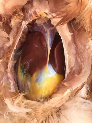
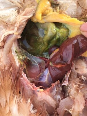
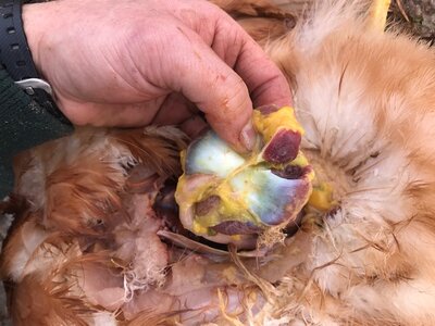
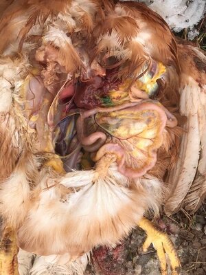
I had a chicken that had signs of Marek's disease. Over the span of 2 weeks she went from good appetite and one paralyzed leg to both legs acting paralyzed and barely able to function. By the end of 2 weeks she flopped around helplessly. She tried to eat, but could barely manage a couple of pecks at her food. We culled her today and I did a quick field necropsy with a dull knife. I have a couple questions about what I saw and attached some photos here. There was a bright green fluid - is that normal? There was a large structure, donut shaped, in her belly. I cut it, and it was a uniform tissue. What was that? Do her follicles look ok? I probably didn't find any diagnostic features of Marek's, but I'd sure like another opinion.
 

Attachments

  • IMG_1330.jpg
    IMG_1330.jpg
    67.6 KB · Views: 17
  • IMG_1335.jpg
    IMG_1335.jpg
    75.6 KB · Views: 17
  • IMG_1337.jpg
    IMG_1337.jpg
    95 KB · Views: 16
  • IMG_1358.JPG
    IMG_1358.JPG
    89.9 KB · Views: 17
I don't know the answers to your questions...but I wanted to offer a suggestion. I had a hen who died this spring after being sick from salpingitis. Hubby and I did a necropsy on her and filmed it, took pictures, etc. I sent the video and pictures to one of the professors at University of Arkansas who works with poultry. He was gracious enough to look over my pictures and video and give me a good diagnosis (salpingitis) of what killed my girl. You might want to try this option? You could either try U of A or one of the many other colleges that specialize in Animal Science...
 
Thank you. It's too late to make a video for this chicken, but maybe I will if more die. Looking at more photos, I'm pretty sure the donut shaped organ was the gizzard - it just wasn't where I expected it to be.


Lots of really smart people on here. I'm sure they will comment soon....

But in the meantime, this is a free online course about chickens. There is a necropsy video in the course that might be helpful. I watched it several times before doing my own necropsy on my girl.

https://courses.uaex.edu/course/index.php?categoryid=76
 
The liver looked super clean to me. I believe I must have nicked the gallbladder, though I didn't notice when I did. It was amazingly bright green I must say.
 
The organ that you're asking about, the bulbous one, is the gizzard. Looks like you nicked the gallblader (bright green bile); that organ is attached to the liver. Appears that this was an unstarted pullet, I see immature yolks, but none that appear ready to become eggs.
Nothing strikes me as out of place or unusual about her internal organs.
 
Thank you. It's too late to make a video for this chicken, but maybe I will if more die. Looking at more photos, I'm pretty sure the donut shaped organ was the gizzard - it just wasn't where I expected it to be.
 
Sorry for your loss. I am not that experienced with knowing what I am looking at in necropsies. The second picture with the green looks like there could be small tumors or infection in the mucosa, and the green color must be bile. Did you nick the gallbladder by any chance? If not, I don’t know what might have caused that. How did the liver look to you . Did it have any spots?
 

New posts New threads Active threads

Back
Top Bottom